
Julia Fiegler-Rudol – Year 9 Student
Julia Fiegler-Rudol (Year 9) writes on the complex topic of the medical application of bioluminescence, in response to the recent Write a Science Blog competition advertised in The GSAL Journal. This excellent essay demonstrates Julia’s passion and curiosity in a field that is pushing the boundaries of 21st century medicine. CPD
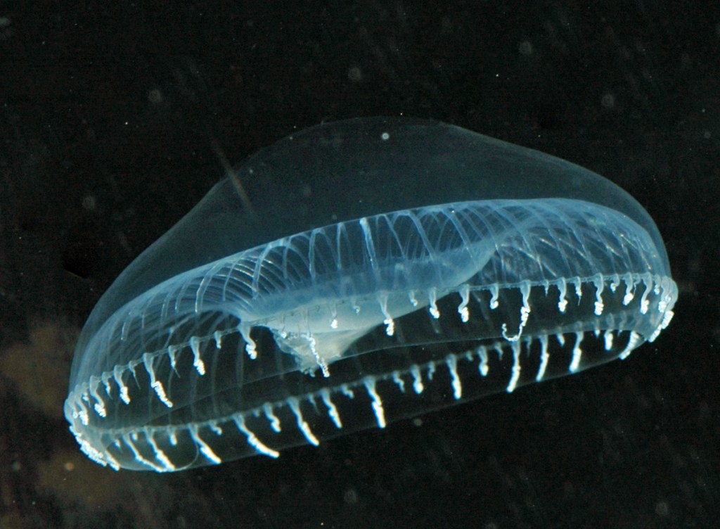
Certain species from the animal kingdom have possessed the ability to present themselves… glowing in the dark. This feature is called bioluminescence – literally meaning production of light by a living creature. It is made possible by the function of a highly specialised protein called green fluorescent protein – or GFP for short.
One particular species of a jellyfish has been in the spotlight due to the high concentration of this substance. The bright and glowing beauties of the sea have now been examined and the mechanism of their ‘lantern capabilities’ explained. As expected, it is not that simple…
The light is emitted or produced as the result of interaction of GFP and another protein called aequorin. The latter occurs naturally and was first discovered in the species of jellyfish called Aequorea victoria, hence the name given to the protein. When the reaction takes place in the presence of calcium ions, the mind blowing and previously unexplained blue light is emitted – making the sea waters look similar to the northern lights. This phenomenally striking appearance has raised fear in the hearts of many laymen for decades and such a mentality might have delayed thorough investigation of this interesting yet vital phenomenon.
Bioluminescence research conquering the world
It all started in 1994 when neurobiologist Martin Chalife conducted the pioneering procedure with GFP by inserting it into various organisms.
In order for this protein to be naturally created by the organism, the gene responsible for its synthesis has to be ‘implanted’ into its genome. At first the commonly used Escherichia coli bacterium was used in the front line. And… bingo! It was a success as the transformed bug cells managed to emit green light once exposed to the ultraviolet wavelength; only the cells with a functional GFP were glowing. The research didn’t stop here and many other organisms like protozoa, plants, and fish were further ‘injected’ with the GFP.
Now, what did this change in the medical world? At first it may not sound very exciting because what other than looking green and pretty does GFP-producing E. coli do? In fact, there is a lot of useful information that has been derived from these experiments.
As mentioned, different cells of many different species may successfully produce their own GFP after inserting this gene. The most ‘straightforward’ examples of its use were utilised during investigation into mammal reproduction or… spread of unwanted bacteria. The cellular function could be investigated to a greater depth while new ways of controlling infections have been developed as a result as well.
HIV
With the success of this research in bacteria, the researchers decided to test their luck in virology. They continued by injecting a lentivirus engineered with GFP genes into an unfertilised cat egg. Once this was done the researchers could examine and observe the characteristics of the virus and track its spread in the egg. As a result of the fact that the cat egg has similar characteristics to the human cell, it was used as a model showing what the virus is capable of – with a large amount of attention aimed at identifying its weak spots… ‘what could stop its destructive path?’. This experiment was revolutionary in our understanding of viral infections, including HIV. With such knowledge, modern ways of treating this infection could be modeled – changing dramatically the treatment of AIDS.
The tiny world of neurons
Individual neurons are impossible to see – the majority of us will certainly agree… wrong! Now, they can be visible. Bioluminescence has enabled neurologists to see the previously impossible: a singular neuron cell. By enlightening the neurons the researchers received a tool to improve understanding of their function and behaviour – especially in the context of understanding the number of devastating diseases and development of new treatment strategies conquering them.
Blood clots killing humans are in the past now
Blood clots used to silently ‘murder’ as many as 100,000-300,000 people in the UK every year! But, what if this could be avoided? Once again the research on bioluminescence came with an answer. Luciferase is the GFP from a firefly – similar to that from the jellyfish. It was used as an imaging agent by scientists to observe the pattern of action of heparin – a drug used to thin blood. On this occasion, nearly infrared light was looked for (the wavelength used among others in the night vision equipment by the military). This enabled detection of tiny amounts of blood clotting cascade protein called factor Xa to better monitor the effectiveness of heparin treatment.
Mothers and fireflies
A mammal foetus is connected with a mother’s circulation via its umbilical cord and placenta. The maternal and fetal circulatory systems are not connected though. The junction between the two is a form of biological barrier allowing for a bidirectional transfer or exchange of substances, allowing for the baby to be fed and have the harmful metabolites cleared. The exact details of this mechanism are not fully understood, which potentially hampers development of prenatal treatment methods. Research from bioluminescence imaging with a firefly luciferase on pregnant mice gave a better understanding of the blood-placenta barrier and how well the foetus is protected against the substances ingested by the mother. Similar methods are being used to better understand the blood-brain barrier which controls which molecules can enter or exit the brain’s vascular system – this knowledge will likely help develop new treatment methods in conditions like Alzheimer’s disease or multiple sclerosis.
There is hardly any doubt that bioluminescence derived from jellyfish or a firefly has come a long way into medical science since it was introduced into the bacterial cell back in 1994. From an extraordinary aquarium exhibit and a unique feature of the sea’s life, the jellyfish is conquering the medical world and changing the lives of the many. The very next time you see one on the seashore you may think that such a simple life form has transformed modern medicine. Julia Fiegler-Rudol (Year 9)
References
References are not currently available for this article.

One thought on “Medical Application of Bioluminescence”