
Gopika N – Year 11 Student
Editor’s Note: Gopika N in Year 11 looks at the benefits and consequences of AI in medicine, specifying in Radiology. This essay is very informative and well researched, and considers all the possibilities that AI has to offer. EB
This article explores the current applications of artificial intelligence in radiology as well as the challenges this poses and future opportunities. Artificial intelligence is defined as the capacity of machines to emulate the intellectual functions of humans. Machine learning is a field of AI and computer science that enables computers to learn without being explicitly programmed, and machine learning algorithms improve the more data it is exposed to. A subset of machine learning is deep learning, when a computer has many layers of interconnected algorithms. Deep learning relies on deep neural networks.
These algorithms form large artificial neural networks, which are composed of nodes connected by links. This is comparable to the network of neurons in the brain connected by axons. Artificial neural networks need to be ‘trained’ in order for the network to ‘learn’. After multiple rounds of training on different datasets, the performance of the network improves gradually and then it can be used in clinical practice. The more layers of algorithms and the more training, the higher performance of the network.
The rapid development of AI in radiology has been driven by a need for greater efficacy and clinical efficiency. There has been a dramatic increase in the acquisition of radiological images such as radiographs, ultrasound, CT and MRI examinations. The data from all these examinations grows at a disproportionate rate compared to the number of trained radiologists; consequently, this has caused an enormous rise in their workload. Radiologists, being human, do make errors of perception and decision-making especially when working under time constraints. AI –when working in conjunction with human radiologists- helps to achieve a reduction in error rate with improved efficiency and minimal radiologist input.
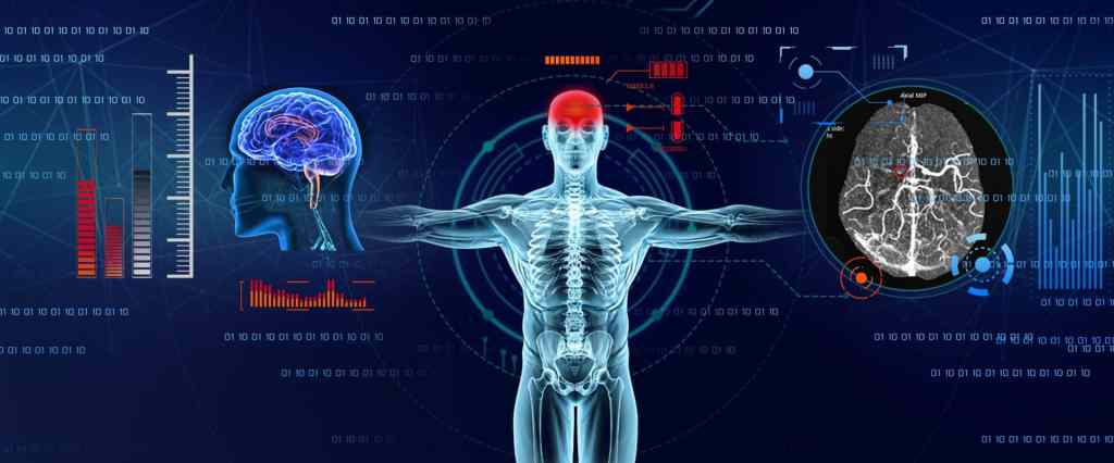
Some general uses of AI in radiology include disease detection and classification, lesion segmentation and treatment response. Additionally, very specific recent applications include identifying lung nodules in chest imaging and also categorizing them as benign or cancerous. AI is able to review large numbers of lung CT examinations and can therefore help with lung cancer screening. Similarly, in abdominal and pelvic imaging, AI can characterise incidental liver lesions detected on CT and MRI as benign or malignant. In Colonoscopy AI can help detect and monitor the growth on colonic polyps that pose a risk of colorectal cancer. AI in mammography can help in breast cancer detection by identifying microcalcifications and tiny lesions that would be incredibly small and difficult to spot with the naked eye. Furthermore, in brain imaging, AI can help in characterisation of brain tumours as benign or malignant and even whether they are primary or metastatic.
However, there are equally many challenges that need to be overcome in order for more effective integration of AI systems. One disadvantage is that currently, AI tools can only address one task at a time however as these systems develop in the forthcoming years, this disadvantage can be easily solved. Another main disadvantage is that to fully automate clinical tasks may take a few years or even decades with current technology, as curating and verifying data for AI is extremely time-consuming. Therefore, complications begin to arise with new diseases that do not have any pre-existing algorithms. Consequently, human readers are almost always required to verify accuracy.
When AI is used in image interpretation, it should be subject to the same stringent regulation as any other medical device. In the USA, it is the FDA and in Europe, it is EU Medical Device Regulation. There are significant legal issues that arise with the use of AI in radiology especially the question of responsibility for an incorrect diagnosis.
There are many possible future applications for AI such as Radiomics – which refers to the extraction of a quantitative value from diagnostic images. This can be used to predict prognosis and treatment response. An interesting prospect for AI is the ability to use large amounts of data to identity imaging markers. For example, in neuroradiology, AI has detected invisible features from MRI pixel data that are not visible to humans. This can predict the genomic composition of brain tumours with greater accuracy than neuroradiologists.
The interesting question is whether AI can replace human radiologists. However, this report believes this is unlikely. It is more probable that the working pattern of radiologists would be enhanced by AI – in fact, radiology work flow is likely to be made faster through the use of AI algorithms.
References:
https://insightsimaging.springeropen.com/articles/10.1186/s13244-019-0738-2
https://pubs.rsna.org/doi/full/10.1148/ryai.2021210118
https://link.springer.com/article/10.1007/s00330-022-08784-6
